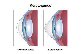
What is keratoconus
.it is a disease that that occurs when the normal round
cornea which known as front part of eye becomes week thin and in regular in
shaped in this disease light does not focused correctly on the retina and
causes disorder of vision
In
the earliest age cut corners causes eye blurring and distortion of eye and
increase gradually to glare the symptoms usually appears in teenage or late
after 20s. Its progress is slow but sometimes it progress rate will be increasesd.As
keratoconus progress the cornea of eye bulge outside and vision become more
weak or blurry.
Eyeglasses
or soft contact lenses are used if the disease is mild stage. As it progress
cornea become thick and changed the shape of cornea.
Keratoconus
does not cause to complete eye blindness but it degrades the level of vision
leading a normal life.
Currently
keratoconus is not 100% cureable.but most cases keratoconus can be successfully
managed. For mild or moderate different types of lenses are used.
·
Most common that helps to detect
it.
·
Near-sightedness
·
Problem in eye focusing
·
Disported vision
·
Blurry vision
·
Light sensitiv
·
Why does keratoconus
happen?
Radiation
exposure
Excessive
eye rubbing
Poorly
or bad fitted contact lenses
Genetic
predisposition
·
Treatments for progression
of keratoconus
·
Corneal cross linking (CXL)
·
Soft contact lenses
·
Gas permeable contact lenses
·
Hybrid contact lenses
·
Sclera and semi-sclera lenses
·
Prosthetic contact lenses
·
Keratoplasty
·
Corneal transplant
Corneal Cross-Linking
methodology to Cure Keratoconus:
This surgical methodology is solely supported the appliance
of a thick answer of lacto Flavin unremarkably referred to as B-complex vitamin
to the attention and exposure to controlled ultraviolet radiations for about
thirty or fewer minutes. The Ribolavin is applied to causes new bond formation
among albuminoidal bands of a tissue layer stromal layer. New bond formation
offers strength and thickness to the fragile membrane that got skinny before
and additionally helps to urge its original spherical structure back. This is
often be} a customary protocol however surgeons can love in 2 ways that. The 2
basic varieties of tissue layer cross-linking area unit mentioned here;
Epithelium-off CXL:
In this sort of cross-linking methodology skinny outer layer
of the membrane is removed. This removal is completed to permit a lot of
penetration of the liquid Ribolavin in order that it will reach to all or any
cells to recover injury.
Also Read: four varieties of Complaints that may be resolved
by optical device Cataract Surgery
Epithelium-on CXL:
This is additionally named because the Trans epithelial
procedure. during this kind, the protecting tissue layer animal tissue isn't
removed however left intact. during this case, loading time of lactoflavin
multiplied in order that it properly reaches to the cells to recover all
injury.
Advantages of tissue layer Cross-Linking Technique:
Corneal cross-linking is that the methodology of alternative
among several different strategies to cure astigmia. it's claimed higher thanks
to following reasons;
It is approved and safe thanks to cure the abnormality
Success rates area unit quite satisfactory
Patients of all age teams even the older one will get
advantage of this surgical cure
It straight off correct the tissue layer injury
It has no semipermanent facet effects
It is quite safe for pregnant and fresh mothers in a very
long haul.


No comments:
Post a Comment